The Science of Good & Evil
(abridged audio presentation)
Broad in scope, deep in analysis, and controversial. Is it human nature to be selfish or selfless, fierce or loving, moral or immoral? Shermer examines the scientific evidence that shows that morality is deeply embedded in our being and behavior. Covers pre-moral animal behavior, neuroscience, game theory, free will, and more.
In this week’s eSkeptic, Mark Perakh discusses how Intelligent Design proponents created the myth that bacterial flagella look like man-made machines.
Mark Perakh is a native of Ukraine. He is a professor of physics (emeritus) at California State University Fullerton. Before coming to the USA as a visiting scientist at the IBM Research Center, he worked for over a half a century as a researcher and university professor in four countries. He has to his credit about 300 scientific papers, four books and several patents. He was awarded several prizes for his research, including one from the Royal Society of London. He has also been active in debunking various versions of crank science. His latest book is Unintelligent Design (2004).
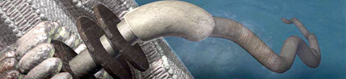
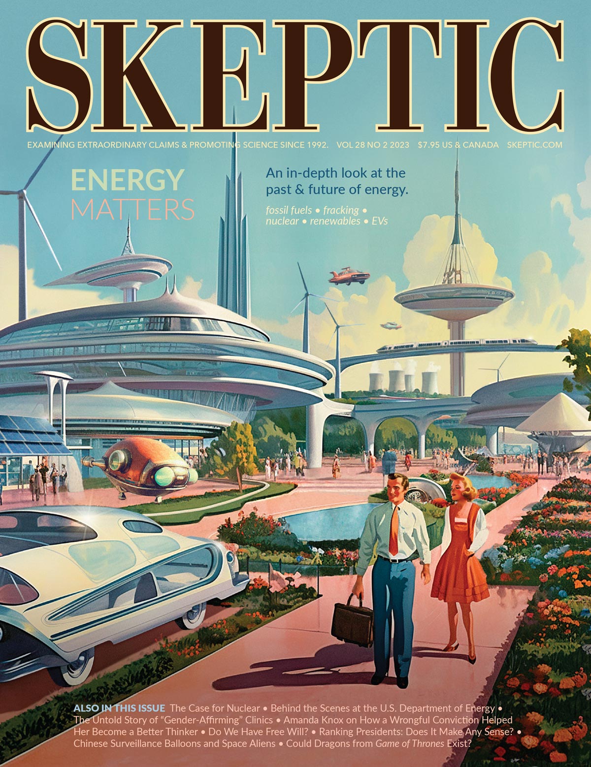
Figure 1: An artist’s rendition of a flagellum as it appears on William Dembski’s blog Uncommon Descent. A similar image appears on the dust cover of Dembski’s book No Free Lunch.
Flagella Myths
by Mark Perakh
In 1996 a professor of biochemistry at Lehigh University named Michael Behe published Darwin’s Black Box1, in which he presented his concept of “irreducible complexity” (IC). Behe and his Intelligent Design (ID) colleagues claim that IC is strong evidence of “design” of biological systems, and ever since his book IC has acquired the status of one of the main pillars of the Intelligent Design platform.
The concept of irreducible complexity was in fact known for many years before Behe’s book. The Nobel Prize winning biologist Hermann J. Müller had already discussed it (under the slightly different name of “interlocking complexity”) in 1918.2 Some 10 years before Behe’s book the same idea was explored by A. Graham Cairns-Smith.3 Unlike Behe, however, these pioneers did not claim that the concept in question was a great discovery on a par with those by “Newton and Einstein, Lavoisier and Schroedinger, Pasteur, and Darwin” (as Behe asserted in Darwin’s Black Box). Neither did they claim that “irreducible complexity” was a “marker” of a supernatural design. To the contrary, according to Müller, development of interlocking complexity in biological systems is to be expected from Darwinian evolution. Therefore the concept in question, as such, evoked no resistance from mainstream science.
As an example of an allegedly irreducibly complex system Behe suggested a mousetrap. Soon afterwards, in multiple publications by various Intelligent Design advocates, images of a mousetrap were endlessly reproduced. The mousetrap, however, was not accepted by the mainstream scientific community as a genuine example of IC. For example, professor of biology John McDonald suggested4 an animated illustration of how, starting with just a piece of a hook-shaped wire serving as a primitive mice-catching device, a full-fledged mousetrap can be gradually built up via two-part, then three-part, etc. contraptions, improving its mice-catching ability at each step in a Darwinian fashion.
Apparently finally realizing that a mousetrap was not a very successful choice for illustrating Behe’s IC concept, ID advocates switched to another example — a bacterial flagellum, a “device” used by bacteria for motility.5 By 2002, the image of a flagellum had become a ubiquitous accompaniment to ID advocates’ books, papers, lecture slides, etc. According to one of the main advocates of ID, William Dembski, the flagellum had become the “mascot” of ID. The image of the flagellum appeared on the cover of Dembski’s book No Free Lunch6, on creationist blogs, etc. Figure 1 (above article title) shows an image of a flagellum as it appears on Dembski’s blog named Uncommon Descent. Notice the smooth surface of the depicted contraption, its perfect symmetry, its tightly fitting components — features we usually see in man-made machinery. This image is a product of an artist’s imagination of how a flagellum “must” look. Does this image truthfully represent the real flagellum? No.
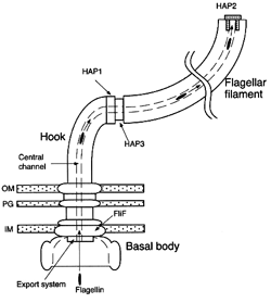
Figure 2: A schematic model of a flagellum. From Yonekura, K., S. Maki, D. G. Morgan, D. J. DeRosier, F.Vonderviszt, K.Imada, and K. Namba, 2000. “The Bacterial Flagellar Cap as the Rotary Promoter of Flagellin Self-Assembly”, Science 290: 2148–2152.
Flagella are tiny organelles that can’t be seen directly by the unaided human eye. Their dimensions are measured in nanometers (billionths of a meter). Modern versions of cryogenic electron microscopy and of X-ray techniques have, though, enabled scientists to form a pretty good understanding of flagellum’s structure and shape. Figure 2 shows a schematic model of a flagellum’s structure.7 This model (one of several published in scientific literature) is a theoretical interpretation of the data obtained via electron microscopy, and mainstream scientists construe it more as an idealized schematic than a true-to-life representation of a flagellum’s actual structure. ID advocates, however, happily treat such images as if they are real replicas of the tiny flagella, usually providing no disclaimers as to the degree of idealization inherent in such images.
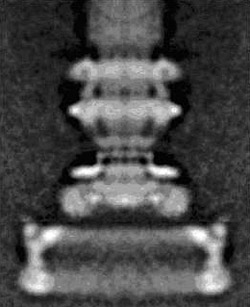
Figure 3: A composite electron micrograph of flagella. It has been obtained by a superposition of multiple photographs shot from various angles, of a number of flagella. From Francis, N. R., Sosinsky, G. E., Thomas, D. and DeRosier, D. J., 1994. “Isolation, characterization and structure of bacterial flagellar motors containing the switch complex.” J Mol Biol. 235 (4), 1261–1270.
In 2004, when Dembski debated Professor Niall Shanks (at UCLA) he displayed8 a different image of a flagellum (Figure 3). Unlike Figures 1 and 2, Figure 3 is neither an artist’s rendition, nor a schematic theoretical model; it is a “real” electron-microscopic photographically obtained image. While produced by scientists, such images are often exploited by ID advocates who are fond of pointing out their striking similarity to man-made machines. However, such illustrations are misleading, picturing the flagellum in a geometrically perfect shape, fully symmetric and consisting of geometrically perfectly formed parts. The real flagellum is far from having such a perfect geometric shape. Unlike machines, which may be close replicas of each other (say, all Jeeps of the same year have almost exactly the same shape) the real flagella, first, have shapes with many deviations from a perfect geometric symmetry, and, second, there are no two flagella exactly identical. Individual flagella differ in various respects, just as biological organisms vary from individual to individual.
Likewise, when I debated Behe on February 15, 2008 on a Larry Kane’s TV show on the Comcast network, Behe supported his pro-ID thesis by displaying the same images of a flagellum as shown in Figures 1, 2 and 3. Behe’s argument essentially boiled down to the stale asseveration that can be succinctly summarized as follows: “You see — it looks like a man-made machine! If it looks like a duck, then it must be a duck! All machines we are familiar with have been designed. Therefore the flagellum must be a product of intelligent design!” Oddly, Behe and his ID friends seem not to realize that the “it must be a duck” argument is an obvious non-sequitur: there are numerous examples of objects whose appearance is deceptive. Just think of the mimicry, so common in nature. For example, look up the article on “Mimicry” in Wikipedia where examples are presented of animals looking like “twigs, bark, leaves, or flowers” etc., thus negating the “it must be a duck” conclusion.
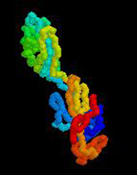
Figure 4: The structure of the hook—a part of a flagellum. From Samatey F.A., Matsunami, H., Imada, K., Nagashima, S., Shaikh, T.R., Thomas, D.R., Chen, J.Z., Derosier, D.J., Kitao, A., Namba, K. “Structure of the bacterial flagellar hook and implication for the molecular universal joint mechanism.” Nature. 2004, Oct 28;431(7012):1047. (In the online version the image is animated, illustrating the flagellar hook’s rotation, see this webpage. Reproduced in accordance with the blanket permission granted in the referenced website, stipulating that a reference to the above article as well as to proteinexplorer.org is provided.
In fact the images that Behe, Dembski, and their ID colleagues show are often not pictures of real flagella. Some of them are just products of an artist’s imagination (Figure 1); others are computer-generated images of imaginary machine-like contraptions. The schematics like that in Figure 2, while reflecting many actual features of flagella, are products of a modeling approximation which likewise can’t pretend to reflect adequately the actual structure of a tiny organelle. However, some other pictures of flagella may indeed be “real” photographically obtained images (Figure 3). Are the images in the latter category adequate representations of the flagella structure?
Look again at Figure 3. It is, at a glance, impressive. Indeed we see here a contraption which is symmetric, its structure machine-like, so it is easy to understand the satisfaction of Dembski and Behe at the sight of this contraption so neatly fitting in with their “design” hypothesis. There are, however, two important details that must be noted. The first detail is that the image in Figure 3 is a composite photo. It is the result of a superposition of many photos, of several flagella, made from various angles. This way the image in question is eliminating from view various imperfections which, unlike in man-made machinery, are inherent in every natural flagellum. Moreover, the procedure of superposition of a number of photos eliminates from view the inevitable individual differences between various flagella, which radically distinguish flagella from “designed” machinery.
The second detail is that the resolution of this picture is insufficient to see the flagellum’s intrinsic structure. To appreciate the significance of this, recall, by analogy, the “face on Mars,” or Lowell’s nonexisting Martian “canals.” When the resolution is insufficient, we “see” nonexisting structures, which on closer inspection look dissolve into natural patterns. This is equally true for the images of very small objects perused under insufficient magnification and/or resolution. The images of flagella obtained at higher resolution, and assisted by other modern sophisticated methods of investigation, reveal the actual configuration of flagella, demonstrating that the seeming machine-like appearance of the flagella in Figure 3 is deceptive.
Figure 5: The structure of the flagellar filament. Side views showing the inner side (left) and outer surface (right). The amino acid sequence of each flagellin subunit is color-coded. From Yonekura, K., Maki-Yonekura, S., Namba, K. “Complete atomic model of the bacterial flagellar filament by electron cryomicroscopy.” Nature, 424: 643–650 (2003); See the online version.
It should be noted that scientists often use such terms as “machine” when describing various biological assemblies. This usage, however, unlike in case of ID advocates, is purely metaphorical, reflecting the superficial resemblance of certain biological structures to man-made machinery. Scientists normally do not imply that biological entities are intrinsically similar to man-made machinery. Perhaps such a usage by scientists is not very fortunate given ID advocates’ misuse of the superficial resemblance between the designed man-made objects and natural biological entities. We have to realize, though, that scientists by and large are not aware of ID advocates’ misuse of such terminology, as only a small minority of scientists pay any attention to ID advocates’ actions.
Let us look at a few selected illustrations of my thesis. The detailed images of the flagella structure obtained via cryogenic electron microscopy combined with sophisticated X-rays techniques are exemplified in Figures 4, 5, and 6. These images, showing the actual configuration of the flagellum, have been selected practically at random from numerous similar images available in the scientific literature. Instead of tightly-fit machine-like parts, we see in these pictures convoluted garlands of protein molecules. These structures look similar to typical bacteriophage viruses5, and have nothing in common with any man-made machine. They vividly illustrate that the image shown in Figure 3 is deceptive and owes its machine-like appearance to the insufficient resolution (not to mention the utter artificiality of the artist’s renditions of flagella, whose variations serve as “mascots” of ID).

Figure 6: Partial structure of the flagellar filament’s cross-section. By Keiichi Namba. See this webpage.
ID advocates often point to the allegedly fraudulent “icons of evolution” utilized by “Darwinists” for their nefarious purposes.9 One such “icon” are the illustrations of embryos made by the 19th-century German biologist Ernst Haeckel. In fact, the faults of Haeckel’s embryological illustrations (dated 1874) were revealed not by creationists but rather by the “Darwinists” themselves.10 On the other hand, creationists of various hues, including ID advocates such as Dembski and Behe, incessantly reproduce images of flagella that are often heavily doctored, without any disclaimers as to the great degree of idealization inherent in these images. Indeed, look again at the images of flagella’s actual molecular structure, as shown above in Figures 4, 5, and 6, and it becomes obvious that real natural flagella are far from looking like man-made machines.
An interesting question arises: Why ID advocates and other creationists, who so eagerly and persistently display pictures like those in Figures 1, 2, and 3, never deign to show much more realistic representations of flagella structure such as those shown in Figures 4, 5, and 6? If they are unaware of these better pictures, perhaps they should try to educate themselves regarding the entirety of the available information about flagella? If, though, they are familiar with the images such as those shown in Figures 4, 5, and 6 (which are freely available both in print and on the internet) could it then be that they are less interested in facts and truth and more focused on winning the “cultural war” by any means?
We must conclude that the argument in favor of “design” of biological entities based on their alleged similarity to man-made machinery is not supported by evidence.
Acknowledgment: My thanks to Matt Young, Paul R. Gross, and Nicholas Matzke for pithy advice.
References
- Behe, Michael, 1996. Darwin’s Black Box. New York: Free Press.
- Muller, Hermann J. 1918. “Genetic Variability, Twin Hybrids and Constant Hybrids, in a Case of Balanced Lethal Factors.” Genetics 3: 422–499.
- Cairns-Smith, A. Graham 1986. Seven Clues to the Origin of Life: A Scientific Detective Story. Cambridge University Press.
- MacDonald, John. Online: http://udel.edu/~mcdonald/mousetrap.html, last accessed on June 20, 2008.
- Luria, Salvador E., Stephen J. Gould, and Sam Singer. 1981. A View of Life. Menlo Park, CA: The Benjamin Cummings Publishing Co.
- Dembski, William A. 2002. No Free Lunch: Why Specified Complexity Cannot Be Purchased Without Intelligence. Lanham, MD: Rowman & Littlefield Publishers.
- Yonekura, K., S. Maki, D. G. Morgan, D. J. DeRosier, F. Vonderviszt, K. Imada, and K. Namba, 2000. “The Bacterial Flagellar Cap as the Rotary Promoter of Flagellin Self-Assembly.” Science 290: 2148–2152.
- Perakh, Mark. 2004. “Three SH’s and One D.” Online at Panda’s Thumb: http://pandasthumb.org/archives/2004/06/three-shs-and-o.html ; last accessed on June 20, 2008.
- Wells, Jonathan. 2002. Icons of Evolution: Science or Myth? Why Much of What We Teach About Evolution is Wrong. New York: Regnery.
- Nic Tamzek (Nicholas Matzke). “Icon of Obfuscation”. In Talk Reason: www.talkreason.org/articles/icon.cfm (last accessed on June 20, 2008).